


Understanding
Keratoconus Management
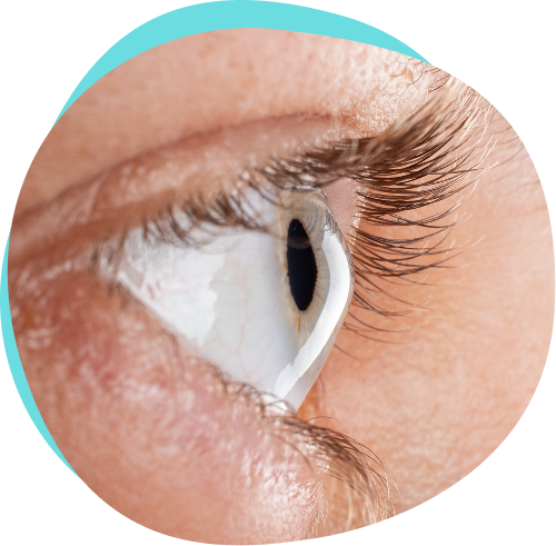
Understanding Keratoconus
asymmetric stromal thinning, cone-like bulging and reduced biomechanical strength, often leading to
significant vision loss.1,2,3
Keratoconus: A Progressive Corneal Ectatic Disorder
The cornea may be affected by a disease known as keratoconus.
Keratoconus is not an inflammatory disease; rather, it is a disorder in which the corneal structure progressively loses rigidity and resistance. As a result, the stromal tissue thins and deforms, primarily in the central region, adopting a cone-like shape, from which the name of the disease itself derives.
It is a chronic and progressive condition, typically bilateral and asymmetric; however, its progression can vary in severity between the two eyes. While historically classified as a non-inflammatory disorder, recent research suggests that inflammatory factors may contribute to its pathogenesis, linking keratoconus to dysregulated immune responses and ocular surface inflammation.
In addition to the classic form of the keratoconus, which we have just described, it is possible to observe an acute form, characterised by the quick, sudden evolution and progression of the phenomenon.
This form is often consequent to a traumatic event.1,2,3

KERATOCONUS: EVOLUTION
1
LEVEL
rEFRACTION
Induced myopia/
astigmatism:
< 5 diopters
cURVATURE
< 48 Diopters
TRANSPARENCY
Corneal scars
PACHYMETRY
> 500 Micron
2
LEVEL
rEFRACTION
Induced myopia/a
stigmatism:
> 5 diopters
< 8 diopters
cURVATURE
< 53 Diopters
TRANSPARENCY
Corneal scars
PACHYMETRY
> 400 Micron
3
LEVEL
rEFRACTION
Induced myopia/a
stigmatism:
> 8 diopters
< 10 diopters
cURVATURE
> 48 Diopters
TRANSPARENCY
Corneal scars
PACHYMETRY
400 – 200 Micron
4
LEVEL
rEFRACTION
Refraction cannot be measured
cURVATURE
> 55 Diopters
TRANSPARENCY
Corneal scars
PACHYMETRY
< 200 Micron

Gaspare Monaco Ophthalmologist
level 1
The first stage takes the name of formed fruste keratoconus and can be confused with simple astigmatism or myopia, as the pachymetry does not show an abnormal corneal thickness, its curvature, in fact does not exceed 48 dioptres.
The exam able to identify it is corneal topography, which allows for mapping of the corneal surface and identifies any suspected irregularities.
level 2
In the second stage of keratoconus, on the other hand, there is an increase in astigmatism and myopia quickly, which requires the correction of the visual defect with constant changes in the ophthalmological prescription.
The corneal curvature, in this case, does not exceed 53 dioptres. Pachymetry, in this case, begins to show a thinner cornea.
level 3
The third stage, given the shape of the cornea, necessarily requires the use of custom semi-rigid contact lenses: the lenses, in fact, are shaped according to the corneal curvature, since glasses can no longer correct the visual defect, and soft lenses are particularly uncomfortable. The corneal curvature is in fact greater than 53 dioptres.
level 4
The fourth stage is characterised by a corneal curvature greater than 55 dioptres, with ectasia visible to the naked eye and extremely reduced corneal thickness.
This represents the most severe stage, for which a donor corneal transplant is envisaged.
KERATOCONUS: ETIOLOGY
& RISK FACTORS5-10
- Sporadic autosomal dominant with reduced penetrance
- First-degree relatives are at much higher risk
- Monozygotic twins show a high concordance of keratoconus
- Preponderance of men over women
- In the UK, Asian patients have a 4x increase in incidence, compared with white patients
The etiopathogenesis of keratoconus is not yet fully known and, as you can see from the slide, there are many possible causal factors.
The genetic factor is associated with 7% of keratoconus cases found, an AD transmission with incomplete penetrance or an AR transmission is hypothesised, in which different genetic subtypes could follow a different mode of transmission.
There is a genetic familiarity that is found between 4 and 23.5% of subjects affected by the disease. A high rate was also observed in monozygotic twins, equal to 71%, and higher than that of dizygotes.
Finally, it is associated with genetic diseases.

KERATOCONUS: ETIOLOGY
& RISK FACTORS5-10
- Contact lenses related injury on keratoconic corneas: release of apoptotic cytokines
- Eye rubbing: 60s of eye rubbing, a 0.5 diopter of astigmatism was found to be induced
- Other diseases: down syndrome, atopic disease, leber congenital amauosis, connective tissue disease and taperetinal degeneration
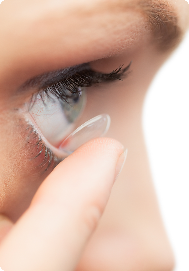
Keratoconus, particularly in its acute form, as we have previously described, is also associated with mild and continuous traumas over time such as that deriving from the use of contact lenses, in particular semi-rigid ones.
The diseases to which keratoconus is associated are both ocular ones, such as retinitis pigmentosa, retinopathy of the premature and allergic keratoconjunctivitis, but it is also often present in subjects with systemic diseases such as osteogenesis imperfecta and Addison’s disease.
Eye rubbing can on the one hand be associated with mild and continuous trauma that encourages the development of keratoconus, and, on the other hand, it is a consequence. In fact, it has also been indicated as a symptom.
Other diseases such as down syndrome, atopic disease, leber congenital amauosis, connective tissue disease and taperetinal degeneration.
Advancing Eye Care Through a Complete Product Portfolio
Evidence-based solutions designed to support clinical excellence
References:
1. Asimellis G, Kaufman EJ. Keratoconus. [Updated 2024 Apr 12]. In: StatPearls [Internet]. Treasure Island (FL): StatPearls Publishing; 2025 Jan-. Available from: https://www.ncbi.nlm.nih.gov/books/NBK470435/
2. Rohit Shetty, et al., Chapter 11 – Inflammation in Keratoconus, Editor(s): Luis Izquierdo, et al., Elsevier, 2023, Pages 159-168,
3. Jacinto Santodomingo-Rubido, et al., Keratoconus: An updated review,Contact Lens and Anterior Eye, Volume 45, Issue 3, 2022, 101559,
4. Gaspare (image)
5. Mas TurV, et al. A reviewof keratoconus: Diagnosis, pathophysiology, and genetics. SurvOphthalmol. 2017;62(6):770-83;
6. AndreanosKD, HashemiK, Petrelli M, et al. KeratoconusTreatment Algorithm. OphthalmolTher. 2017;6(2):245-62;
7. AsimellisG, Kaufman EJ. Keratoconus. StatPearls. TreasureIsland (FL)2018.
8. Ertan A, Muftuoglu O. Keratoconus clinical findings according to different age and gender groups. Cornea. 2008;27(10):1109–1113.
9. Millodot M, Shneor E, Albou S, Atlani E, Gordon-Shaag A. Prevalence and associated factors of keratoconus in jerusalem: a cross-sectional study. Ophthalmic Epidemiology. 2011;18(2):91–97
10. Weed KH, MacEwen CJ, Giles T, Low J, McGhee CNJ. The Dundee University Scottish Keratoconus study: demographics, corneal signs, associated diseases, and eye rubbing. Eye. 2008;22(4):534–541.